Our product
ProtonVDA pRAD System
ProtonVDA team has developed a full proton radiography system (pRAD) with significant clinical capabilities.
The ProtonVDA pRAD System is a proton imaging system that uses tracking detectors to measure the transverse position of individual protons before and after the patient, and a residual range detector to determine the proton energy absorbed within the patient.
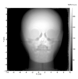
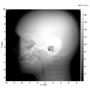
Our detectors produce an image from position and energy measurements of protons with enough energy to traverse the patient.
Our advantage lies in our insight that proton imaging can be fast, light-weight, and seamlessly integrated into proton therapy systems and clinical workflow.
Examples of Proton Images
AP view of paediatric head phantom (left). lateral view of pediatric head phantom (right).
Key benefits of pRAD system
Treatment planning
A valuable tool for treatment planning.
Range checks
Suitable for daily range checks and patient alignment.
Low radiation dose
Low absorbed dose required for a pRAD scan of a head (10 times less than X-Rays). Imaging can be done for each fraction and could be used to detect anatomical changes.
Proton-beam’s eye view
Proton images can be taken under the same geometrical conditions as the treatment, so the images are from a true proton-beam’s-eye view.
Accurate image of metallic implants
Images free of artifacts from high-Z implants
The ProtonVDA pRAD System has a setup at the Northwestern Medicine Chicago Proton Center and is currently beeing medically certified.
pRAD System’s detectors and Image Reconstruction Software is designed with Proton Computed Tomography (pCT) upgrades in mind. The use of proton computed tomography (pCT) will add the capability of obtaining a directly measured 3D proton stopping power map for use in treatment planning.
Download
Proton Radiograph (pRAD System) brochure
Would you like more information on the ProtonVDA pRAD System?
Download the brochure
Start
a Dialog
with Us


