A Comparison of Proton Stopping Power Measured with Proton CT and X-Ray CT in Fresh Post-Mortem Porcine Structures.
Don F. DeJongh, Ethan A. DeJongh, Victor Rykalin, Greg DeFillippo, Mark Pankuch, Andrew W. Best, George Coutrakon, Kirk L. Duffin, Nicholas T. Karonis, Caesar E. Ordonez, Christina Sarosiek, Reinhard W. Schulte, John R. Winans, Alec M. Block, Courtney L. Hentz and James S. Welsh
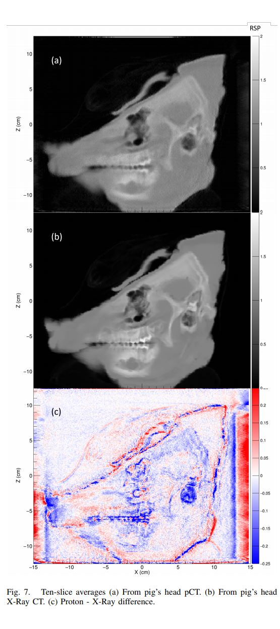
Abstract – Currently, calculations of proton range in proton therapy patients are based on a conversion of CT Hounsfield Units of patient tissues to proton relative stopping power. Uncertainties in this conversion necessitate larger proximal and distal planned target volume margins. Proton CT can potentially reduce these uncertainties by directly measuring proton stopping power. We have used a prototype proton imaging system to acquire proton radiography and proton CT images of a sample of pork shoulder and ribs, as well as of a pig’s head. Close in time, we also acquired X-Ray CT scans, and compared proton stopping power measurements from the two modalities. We find agreement within 1% to 2% for soft tissues, and discrepancies of up to 6% for compact bone. We observe the largest discrepancies, up to 30%, for cavitated regions with mixed content of air, soft tissue, and bone, such as sinus cavities or tympanic bullae. If these results apply in general, it may be possible to substantially reduce uncertainty margins for many treatment plans, while avoiding regions with higher uncertainties. Our comparison of proton radiography data with a digitally reconstructed radiograph from the X-Ray CT demonstrates the potential of this modality for daily pre-treatment range verification.